Social Interaction
Video-Based Studies of Human Sociality
Toward a praxeological account of performing surgery:
Overcoming methodological and technical constraints
Satomi Kuroshima1 & Jonas Ivarsson2
1Tamagawa University
2University of Gothenburg
Abstract
Surgical operations are fundamentally comprised of multisensorial and multimodal activities. As surgical work involves professional and technical skills that entail a multitude of sensorial information, various methodological difficulties and technical constraints emerge for analysts. Subjective sensations and feedback received during the participants' constructed actions may not be available to outsiders, and the privilege of studying surgical operations is not always guaranteed for the fieldworker. However, as practical surgical tasks are constructed from the routine progression of mundane activities, technical and methodological difficulties can be overcome, confirming the perspicuous nature of surgical operations for social scientists as outsiders. In this report, the researchers describe their fieldwork experiences in two different types of operating rooms—gastroenterology surgical operations in a Japanese context and endovascular aortic repairs in a Swedish context—with a specific focus on how they controlled the technical challenges. This demonstrates the value of surgical operations as a site for scientific investigation independent of expert knowledge about surgery.
Keywords: multisensoriality, multimodality, practical reasoning, sensation, touch, perception, knowledge
1. Introduction
Given surgery’s business of performing physical interventions in patients’ bodies, it consists largely of multisensorial and multimodal activities. For instance, tactile perceptions of the tension encountered when using equipment are critical for enabling the precise calibration of a piece of surgical equipment for an operation; feedback pressure from human organs can provide important signs of whether a tumor has developed or not. Other sensory resources, such as smell, also inform the participants that a particular procedure (e.g., tissue burning) is ongoing. Even with contemporary technologies to aid and augment the human capability to perform surgery (e.g., the da Vinci surgical robot), in many cases, surgical practice still relies on a human’s keen sense to grasp the physical condition of the patient’s body and the mastery of natural language (Garfinkel & Sacks, 1970) to accountably know what each member of the activity is doing at any particular moment. Furthermore, within such a specific activity framework as surgery, the participants selectively perceive available sensorial information to accomplish activity-relevant actions. As in any multimodal setting, the medical staff strive to understand “Why that now?” through the fields of action opened by complex contextual configurations (Goodwin, 2000). Given the bearing of a range of sense perceptions on understanding action, we should consider multisensoriality a subject for further scientific investigation (Mondada, 2019).
Nevertheless, technical and methodological difficulties abound for capturing multisensoriality in medical and surgical settings where technical instruments and procedures fundamentally structure the interaction among practitioners, and where there is minimal tolerance for interference (e.g., due to hygienic concerns). On the one hand, some challenges manifest as physical restrictions for conducting data collection, for example, when a researcher is not allowed to freely position cameras and microphones during fieldwork. Operating rooms are inevitably full of noise from various kinds of machines and ventilation systems, which can also hinder the camera’s view. Not unexpectedly, the operating rooms’ physical conditions often restrict research documentation. On the other hand, surgical operations are highly sensitive, technological, and technical settings which make them exacting objects of study (see Koschmann et al., 2007; Koschmann et al., 2011; Mondada, 2011, 2014a, 2014b; Zemel & Koschmann, 2014). Sometimes, the analysis of domain-specific phenomena requires a level of understanding that almost matches that of the research subjects. One way out of this problem (besides becoming a surgeon) is to see the scene through the eyes of the participants and closely follow their practical reasoning as to how they make sense of one another (Garfinkel, 1967). After all, within a situated activity, we understand perception as intersubjectively structured rather than as a subjective or private phenomenon. In this way, praxeological accounts of participants’ conduct—accounts that outline the ways in which actions are reflexively tied in with the practices they help shape—also become a way to hone in on the multisensoriality of human activity. Building on this, we nurture an interest in domain-specific phenomena and tasks, as situated accomplishments of parties to a setting. Following Zimmerman and Pollner’s (1970) reasoning, we understand these accomplishments, unique as they are to the settings studied, to still rely on some practices through which those phenomena are made observable. The focus on site-specific work can reveal something of these practices’ invariant properties that operate across settings.
With the above-mentioned analytic goal in mind, we will discuss how to deal with the technical and methodological challenges and constraints of conducting research in medical and surgical settings. This study concerns professional multisensorial interaction during surgical operations and juxtaposes two surgical settings with different technical conditions. As the perceptions and sensations of individuals are regularly made constitutive of particular actions in public settings, both co-present participants and analysts will gain adequate access to the participants’ perceptions through their commonsensical understanding of the scene and the activity (Garfinkel, 1967; Goodwin, 1995; Mondada, 2019).
In what follows, we demonstrate how the researchers endeavored to overcome the aforementioned challenges by focusing on the participants’ conduct through an analysis of video recordings of practitioners dealing with practical tasks in situ, both inside and outside the operating room. We also describe how such analyses can be further informed by collaborating with practitioners, making video an important resource for researchers to accommodate “the unique adequacy requirement of methods” (Garfinkel, 2002, p. 175). This methodological policy, recommended in ethnomethodological studies of work, urges the analyst to engage deeply with the research field to develop their competence in the studied objects to get a high level of descriptive precision.
2. Conducting fieldwork for EMCA studies in surgical operations
In this section, we first introduce two sites where each researcher conducted their fieldwork and data collection. Then, we discuss the role of the researcher and how we overcame certain obstacles in collaboration with the practitioners. Finally, in the next section, we present individual data analyses to demonstrate how our studies of the practitioners’ multisensorial conduct became possible. The amount of detail included in the technical and medical specifics of the fieldwork settings (see the sections 2.1-2.3 as well as 3.5) may be more than required for a reader to comprehend the multisensorial analysis. However, it should be noted that for the praxeological account and the analyst’s work leading up to the analysis, this level of understanding is essential. Such technical and medical requirements pertaining to the work constitute a major part of the background expectancies in play in the studied settings. Naturally, these crop up occasionally in the interaction. Therefore, it is recommended to the readers that a firm grasp of the basic but site-specific conditions of the work which participants already share would enable them to deepen their commonsense understanding.
2.1 Establishment of the EMCA research project
One of our study sites is part of an interdisciplinary research project conducted at a department of gastroenterology in a large public university hospital in the greater Tokyo area, Japan. This project was launched with the aim of studying human action conducted within a highly technical and technologically skillful environment. The fieldwork period ran from 2014 to 2016 and recorded 15 cases of gastroenterological surgery, generating a data corpus of 50 hours of video. The operations that were video recorded lasted a minimum of 2 hours, with some cases lasting more than 5 hours.
The data collection at the site was launched after obtaining ethical approval where the researcher was invited to record operations in which a novel 3D surgical navigation system, a 3D image reconstruction of the patient’s internal body organ structures based on scanned images, was used to help the surgeons grasp and predict where the tumors are located in relation to the neighboring blood vessels (Oshiro et al., 2017). Because of the unique technological features of the surgery, the practitioners’ interactions sometimes involved a discussion of using these navigation systems, and the researcher was expected to learn about and gain procedural knowledge of how these technologies work in addition to more general understanding of surgical terminologies. Typical operations involved the partial or entire removal of organs such as livers, pancreases, or gall bladders. Therefore, decision making as to where and how to perform excisions (which is primarily based on the sensorial feedback and information one would receive from the operation) became an omnirelevant problem for the surgeons.
Our second example concerns work performed in what is categorized as a hybrid operating theater in a Swedish hospital. This fairly recent development in operating suites combines minimally invasive surgery (often relying on X-ray guidance) with open surgery. In these setting, surgical team members with differing expertise and different working methods can collaborate in new constellations.
Due to this novelty, the operating suite used for the study, built as a learning facility, was equipped with multiple ceiling-mounted video cameras and the capability to stream all output from computers and imaging equipment to a monitoring room located elsewhere in the hospital. When this facility was first constructed, the staff raised concerns about the possibility of having their work remotely monitored; therefore, a number of procedures were established to create a feeling of safety and control. As a first measure, recording can only ever be initiated from the control room adjacent to the operating room; it cannot be turned on remotely. Additionally, a number of “On Air” signs are mounted within and outside the operating theater, clearly indicating when recording is ongoing.
Another important aspect of this operating room was its ventilation system and how it restricted recording. Additional bodies and equipment present within the surgical suite would reduce the effectiveness of the ventilation. There could, therefore, be hygienic consequences when recording inside the operating room, even with sterilized equipment kept at a distance. As a non-medical specialist, it is difficult to assess how great a risk this presents. Given the option of making remote recordings, this path was chosen for the project.
2.2 Role as a researcher in the fieldwork
In addition to the details of how the researchers undertook the fieldwork at their respective sites, creating and maintaining rapport with the participating surgeons was equally important to conducting research in such highly specialized settings.
In the Japanese case, the project was initiated by surgeons interested in developing state-of-the-art 3D imaging technology for navigation during an operation. The researcher was later invited to participate in the group due to her specialty in conversation analysis. Even though the initiative came from another party, it was important to demonstrate how research using conversation analysis can be conducted effectively and prove valuable in terms of describing ‘the seen but unnoticed’ (Garfinkel, 1967, p. 36) aspects of a surgical operation. Thus, the researcher presented her findings at several medical conferences and joined research meetings with surgeons whenever possible. Because of this position, the researcher was able to consult with surgeons and technicians on the data and technical aspects of the surgical procedures.
As the sole field researcher, the first author managed all the recordings in the Japanese setting. However, if a scheduled surgery could not be attended by the researcher, staff members of the surgical department helped with the recordings. Because the operating room was not large enough for the surgical team as well as an observer, participant observation was not an option. Therefore, during the surgery, the operating room was only visited by the researcher to check on the equipment. This arrangement was possible only because of the researcher’s enduring efforts to create and maintain rapport with all concerned parties.
On the other hand, the Swedish project was set up as a collaboration among an active surgeon, medical physicists responsible for radiation safety at the hospital, and the researchers. By jointly formulating the project’s goals, much of the required legitimacy for conducting the research was formed at this initial stage. In this case, however, additional communication with the hospital’s management team, union representatives, and the staff at large (i.e., the team of anesthesiologists, nurses, and vascular surgeons) was required to establish an understanding of the project’s goals. The requirement to gather informed consent from all parties potentially affected by the study entailed a four-month long process of meetings before any recordings could be initiated.
Hospitals are highly sensitive areas with specific ethical, legal, technical, and hygienic standards or requirements, and a researcher must be trusted not to interfere with medical practices so as to not jeopardize their integrity. Establishing a level of trust in the researcher’s ability to professionally conduct the research is thus paramount. This can be a long process involving constant demonstrations of how one is capable of providing relevant feedback or observations, which reflexively leads to the building of rapport with practitioners.
2.3. Overcoming technical challenges for making video recordings and analyzing surgical interaction data
As the setup for medical surgery usually involves advanced and cutting-edge technologies, conducting fieldwork for EMCA studies imposes significant challenges on the researchers. In addition to adapting to highly skilled settings, the researchers also have to meet the requirements of sterilization and sensitivity to the type of work that they are studying. In this section, we describe how the researchers in each setting dealt with difficulties indigenous to the type of work they were studying.
At the Japanese site, video recordings were typically conducted using two cameras. The cameras were placed diagonally across the room to capture all participants’ fronts (see Figure 1). We set up one camera in a corner and the other on some equipment installed on the opposing wall to minimize any obstruction of the medical team’s movements and to maintain the environment’s sterility. Even though all participants were technically recorded from the front, they often gathered closely together during the operation. The result was that some bodies would occlude those standing on the opposing side of the operating table.
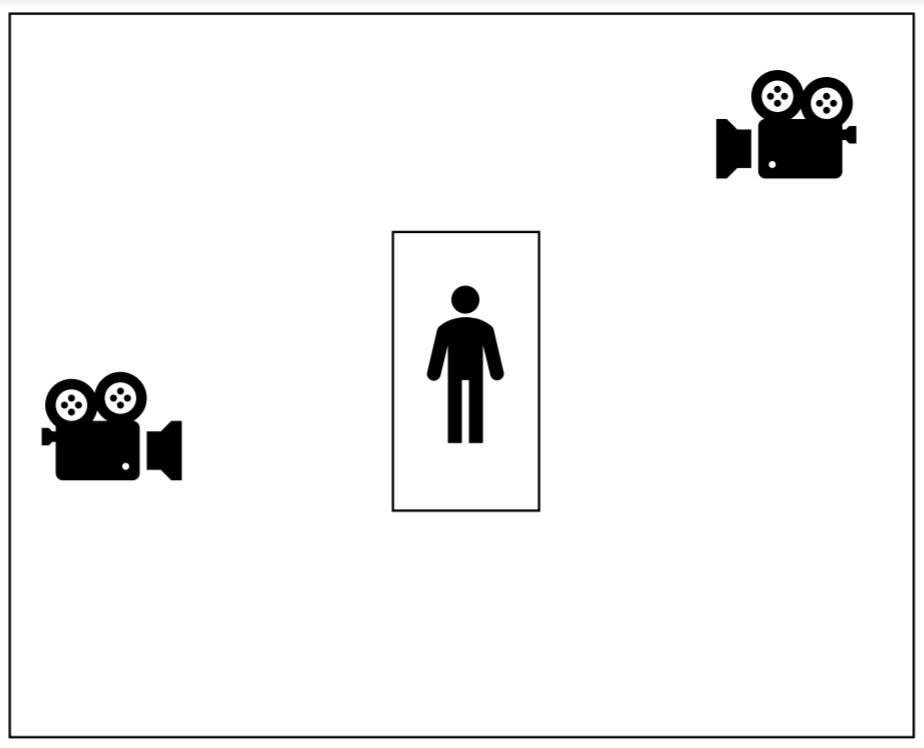
Figure 1.
A central part of the operating room was the surgical monitors displaying the close-up shot of the operation field captured by the operation camera. The monitors hung from the ceiling above the patient’s head. The same footage was available to the researcher after the operation; however, this material did not have any sound. Working from this source made it strenuous to coordinate the images with the conversational data (though it was not impossible). The surgeons declined wearing lapel microphones due to sterility concerns and the possibility that the extra equipment might restrict their movements during operations. For these reasons, wireless microphones were instead mounted on the surgical camera in the operating area, so that the external cameras could record the talk.
Under these conditions, a number of difficulties arose during the subsequent analysis. The noise of the operating room was disruptively loud, with sounds emanating from the various activities, equipment, and people moving about the operating room, and the surgeons’ masks made their conversations difficult to hear. Additionally, on occasion, the equipment, such as the surgical lights and the various types of monitors, interfered with long shots of the scene, making it difficult to see what the surgeons were doing. As a consequence, the researcher had to make the best of the suboptimal recordings. In hindsight, an IC recorder could have compensated for the lack of clear sound. However, at the time, the primary consideration was not to interfere with the surgical field, so no additional recording equipment was introduced.
In the Swedish context, the cameras mounted on the ceiling were of very high quality and could be remotely controlled with respect to pan, tilt, and zoom. A number of XLR connectors for microphones were also spread across the ceiling and on pendants, enabling the selective advance placement of microphones to focus on specific team members. Even so, poor sound quality remained an issue, similar to the Japanese setting. However, with the help of the technicians who had built the sound and video installations, we were able to optimize the sound and noise filtering during the ongoing surgery.
Nevertheless, the sound conditions, even for the participants in the setting, were challenging. The large space combined with the sounds of fans, a large robotic arm carrying an X-ray sensor, and additional noise-generating equipment created a soundscape of varying complexity. At certain times during the recordings, all the voices could be heard clearly, but at other times, particularly when several different groups of staff were talking at the same time, it was difficult to differentiate the separate streams. To counteract this, it would have been helpful to use individual lapel microphones for staff members of interest. However, this would have required wireless microphones. As the operating theater is a technically sensitive setting with respect to the risk of interfering with life-sustaining equipment, as in the Japanese context, we did not explore wireless technology options. Again, we chose to use the existing technical solution despite its potential drawbacks.
For remote viewing unique to the Swedish case, a conference room was fitted with two large monitors and a control panel. Once a session had been initiated from inside the operating room, it was possible to select from among twenty different feeds (e.g., camera, computer, X-ray, ultrasound, and endoscope) and project one feed on each monitor. A major benefit of this setup was that all sound coming from the operating room was automatically integrated with whichever video stream was selected. This had tremendous beneficial consequences for the later stages of synchronizing the sources.
The majority of the surgical work was being carried out in front of a computer monitor hung above the patient, with one surgeon controlling the inserted instruments by hand. To capture as much of the central area of the surgical work as possible, it was decided to use one overhead camera shot in combination with a video stream from the surgeon’s main working monitor. The information displayed on this monitor was found to be essential for following the events and to make sense of the progression of the procedure. These two recordings were then combined into a single timeline using Final Cut Pro editing software. By maintaining the original size of the surgical monitor feed, the same visual access the participants had was available for the secondary analysis.
Another factor when making these recordings was the duration of the surgical procedures. The shortest recordings were two hours long, while the longest recording was over 10 hours long. For the research project, 12 procedures were documented, totaling 58 hours of recording. As a consequence of having very long recordings, Final Cut Pro was found to be the best software for continuing the analysis. Unlike most video playback software, video editing suites typically come with scalable timelines. When this timeline is zoomed out, one can quickly look for events and changes that occur over many hours, and when it is zoomed in, one can identify differences down to individual frames. The added feature of creating markers and other forms of annotation also makes it possible to build a preliminary structure for the analysis into the video materials. In this way, for instance, every occurrence of artificially induced apnea (115 cases) was identified and annotated, and later analyzed and reported (Ivarsson & Åberg, 2020).
Even with this kind of high-quality visual access and the technologically supplemented help, the surgical work and its analysis were not without complications. The selected examples analyzed below in 3.1 illustrate some of the concerns regarding the different sensorial aspects of the studied practices. As we aim to demonstrate, these are concerns, for members of surgical teams as well as for analysts, regarding the accountability of the members’ conduct as the focus of the study—just as in any other perspicuous setting.
3. Observing and accounting for multisensoriality in surgical operations
3.1. Difficulties and challenges in analyzing the multisensorial and multimodal interactions of technical surgical operations
As mentioned, a surgical operation is a multisensorial field. Surgeons dissect, open, and ligate blood vessels and organs. Most of this activity requires the surgeons to “see” various objects and make judgments based on their professional vision (Goodwin, 1996, 1997; Kuroshima, 2018). However, the work also relies on other human sensations such as touch, hearing, and even smell in certain circumstances. For example, when dissecting tissue, tactile feedback from the object is an important resource for a party to assess the possibility of continuing a dissection (see Excerpt 2).
Typically, researchers face serious challenges when analyzing such perception-based actions (Nishizaka, forthcoming). Akin to touch in a professional activity, the subjective experiences mediated via one’s other sense organs are not directly available to other parties in a non-interactional and technical sense. However, as the activity at large cannot be accomplished by just one person’s conduct (Schegloff, 1968), multisensorial aspects of the work have to be brought to the collaboration by the participants themselves. In this way, private perceptions are rendered observable for others as well. Subjective experiences are made available for observation, assessment, and comments to achieve a shared understanding of the surgery at any moment in time.
By making sensations available and analyzable for co-present participants, the subjective experience via one’s sensations is also made available to the researcher observing the activity through a camera lens. For example, professional massage therapists’ tactile perceptions are used as resources to produce not only diagnosis but also the social actions of acknowledging and confirming the problems mentioned by patients (Kuroshima, 2020). Although we have better access to the participants’ visual practice, as we can arguably “see” what they are doing by looking at objects (cf. Gibson, 1966), other senses are not necessarily unavailable as long as they are made public through concerted actions (i.e., the rational accountability of practical activities) (Garfinkel & Sacks, 1970; Nishizaka, 2017; see Streeck’s [2013] discussion about the living body). This point is demonstrated in the following analyses. That is, each analysis shows how the skillful and professional organization of the multisensorial work of surgical operations can be approached. The subsequent analyses, the first three from the Japanese setting and the remaining three from the Swedish setting, are dedicated to describing and analyzing how various types of multisensorial action constitution (e.g., visual, tactile, and auditory perceptions) manifested in interaction. In these praxeological accounts, the common-sense rational properties of multisensorial conduct will be made available. As will be demonstrated, even such distinctly technical activities as surgical operations are made observable through the accountability of practical actions (Garfinkel, 1967).
3.2. Making a judgment based on visual perception
A practical problem for participants in a multi-party activity setting is the establishment of intersubjective ground. One way to deal with this problem is by displaying one’s perceptions in talk. Excerpt 1 illustrates this point. In this segment, the surgical team seems to have reached a critical point for their operation (the partial surgical removal of the liver). At the start of Excerpt 1, the main surgeon (D1) and the two assisting surgeons (D2 and D3) are looking at the operating area. D1 starts making a judgment call to the group (line 2) and subsequently provides an account for doing so (line 4). With his deontic authority of the matter in question—in his position as lead surgeon, he is expected to have the right to propose the next course of action—this judgment is understood as a proposal for the team to prepare to stop (Stevanovic & Peräkylä, 2012). Although the researcher’s camera only captures the surgeons from a distance and their upper bodies (see Figure 2), the surgical area becomes available through the surgical monitors (see Section 2.4 for the arrangement of recording). Though the image of the surgical monitors cannot be quoted in the paper for ethical reason, this view aided the researcher in analyzing the members’ methods of accounting for their visual perceptions as the surgery proceeds.
Excerpt 1.
Open in a separate windowIn line 2, D1’s proposal to stop presents his judgment via his visual perception using deictic terms and ending the turn with a quotation marker -tte, which is used for making a report (Hayashi, 1997). In this way, as akin to Goodwin’s (1996) professional vision, he presents his perspective on the current status of the operation. Despite his deontic authority, he further accounts to justify how his judgment is reasonable by explicating how he sees the blood vessels structure at this point (lines 4 and 5). In particular, he couples his bodily display of how he sees the current status of the operation with the particular setup of the operation, that is, by showing his gaze direction towards the surgical field and pointing at the blood vessel with his scalpel (Figure 2) (Goodwin, 2013, 2018). By producing an action based on his visual perception in this way, D1 invites them to assess the same object, which would establish an intersubjective ground for the current activity (Kuroshima, 2017). The proposal was accounted for most likely not due to the reduced deontic authority, but due to the nature of the proposal which is constructed via a speaker’s perception. Indeed, in line 6, D3 endorses D1’s judgment by repetitional agreement via visibly showing that he is looking at the surgical field; thereby he also constructs his agreement based on his own visual access to the object in question (cf. Nishizaka, forthcoming). After this endorsement is obtained, D1 proceeds to make more specific proposal to discontinue the procedure than the one in line 2 (line 8). During this proposal, D1 shifts his gaze from the operating area to D2 to build a new course of action, which is accepted by D2 in line 10.
In this example, the surgeons’ professional vision becomes available not only to the recipients of these actions but also to laypersons, such as the researcher, through an examination of the accountability of the participants’ practical actions as they are rendered visually accessible to the recipients and sequentially positioned with one another.
3.3. Complaining based on tactile perception
The following excerpt was recorded during a cholecystectomy (i.e., the surgical removal of the gall bladder). D2 is dissecting part of a gall bladder and D1 is watching the work on the monitor showing the laparoscopic image. D2 has been trying to dissect tissue with an electronic scalpel for a while. As a continuing state of incipient talk (Schegloff & Sacks, 1973), D1 remarks that the scene he is observing is noteworthy because of a unique feature of the patient’s organ and seeks agreement from the free participant, D3, by shifting his gaze from the monitor to D3 (line 1). As the prompt to comment on the scene is made, D2, still intensely looking at the monitor (Figure 3), complains about the tissue being especially katai (hard) and that he cannot proceed smoothly.
Excerpt 2.
Open in a separate windowAs a response, D1 agrees with D2 and further validates his complaint by offering an account of why the tissue is hard (line 4) (i.e., because the patient’s organ is inflamed); thus, treating the complaining as reasonable to be initiated at this moment. Although the tactile feedback of the tissue (which is hard) is only received by D2, in this case, D1 is clearly able to endorse D2 by observing how D2 visually presents his problem with the procedure through his description of his tactile perception, body, and manipulation of tools, thus making the scene accountable via a complaining action. This exchange is a clear illustration of how one’s sensorial reception becomes accessible to others based on the formulation of one’s action (i.e., a complaint).
3.4. Sound as a resource for action production
Next, we offer an examination of how an action is constructed and can be made intelligible through its auditory qualities. Excerpt 3 was recorded during a partial hepatectomy (i.e., the surgical removal of the liver). D1 and D2, the main surgeons at this time, have been repeatedly dissecting vessels and stopping the bleeding by using thread to ligate each vessel. Normally, bleeding occurs as they dissect the tissue; therefore, D3 assists the surgery by suctioning the blood while the dissection takes place. However, when the surgeons are ligating, there is no need for suction. At this moment in the excerpt, they have reached a point where a part of the liver has almost been removed. In line 1, D1 requests a thread from the scrub nurse after dissecting a blood vessel. After tying the thread around the blood vessel, D1 minimally marks the completion of the tying action in line 5 so that the next expected action can be occasioned. Indeed, subsequently, D3 starts suctioning the blood in the surgical area without saying anything (line 6). J1 is a resident who is observing the operation.
Excerpt 3.
Open in a separate windowWe can observe by D3’s action that he silently implements the suctioning of the blood in the appropriate and relevant slot within this activity. D1 marks the completion of his tying action in line 5 and, as a result, the next course of action can be initiated. Prompted by this, D3 begins the suctioning. A distinct sound accompanies the manipulation of the suction tube. Coupled with the movement of the suction tube which is visibly available for other participants (i.e., they can see the suction tube; see Figure 4), positioned in this slot without any verbal utterances, such auditory information is an essential resource for constructing this action of suctioning. Without the actual auditory and visual information, the action may not have been comprehensible as suctioning, as the sound is distinctively constitutive of its move. Thus, indeed, even in the absence of a verbal claim that could contribute to its saliency, D3’s action in line 6 is made accountable as performing the suction as the next relevant course of action. Accordingly, the other participants are engaged in other work, such as switching their operation tools, while the suction is ongoing.
These analyses show how the multisensorial construction of practical actions emerges in the surgeons’ activities. Even though the work is both technical and builds on the perceptions of vision, touch and hearing, it is nevertheless made accountable. The current status and progression of the operation are evidenced by these judgements, inviting others to agree by providing complaints or performing timely medical actions such as suctioning.
Furthermore, even if the participants are operating with specialized medical equipment which requires professional training, the specialists can also invite outsiders to share their experiential worlds. The next section demonstrates some methods.
3.5. Technical aspects of the medical procedure
The Swedish recordings focused on an endovascular aortic repair (EVAR) surgical procedure. Used to treat aortic pathology, this procedure places an expandable stent graft with the aid of medical imaging techniques. For percutaneous EVAR, small incisions are made over both femoral arteries in the groin and vascular sheaths are introduced. Guidewires, catheters, and endografts are passed through these sheaths and placed in the correct position using X-ray fluoroscopic guidance. The compressed stent graft is inserted into the patient’s aorta and then expanded in place to provide structural support and alleviate the pressure of the aneurysm on the aortic walls. The deployment of a stent graft is a stepwise procedure. First, its proximal end is expanded by retraction of the delivery sheath, then hooks or barbs are uncovered to penetrate the aortic wall from the inside to permanently fix the stent graft. The main body of a stent graft is sometimes likened to a pair of trousers with one long leg and one short leg. While the longer leg extends down one of the femoral arteries, the shorter leg, also called the contralateral leg, has to be extended by adding a second piece of stent graft introduced from the opposing femoral artery. To enable this docking procedure, a guidewire is introduced from below, where the tip of the wire needs to be navigated through a small opening, or lumen, of the shorter leg.
A problem often encountered at this stage is that the lumen is situated inside the enlarged aorta, a three-dimensional space inside the patient’s body. Physical access to this space is provided by the ports in the patient’s groin. Here, the catheters and wires can only be moved in and out or rotated. As these instruments follow the patients’ (largely hidden) anatomy, there is always an element of uncertainty concerning the results of their manipulation.
The surgeon’s operations are visually guided by X-ray fluoroscopy, a visual modality limited to highly attenuated (i.e., hard) structures. This makes visible the instruments and the structural elements of the stent graft. Except for the spine, most of the patient’s anatomy remains invisible. For this particular task, interactions between the instruments (i.e., the metal wire and the catheter) and the metal components of the stent graft are central.
3.6. The practical problem of understanding touch and tension
One of the things missing for us as onlooking analysts is the tactile sensation of the instruments as they are being manipulated. There are obvious differences in stiffness of the existing varieties of wires and catheters. Furthermore, the insides of arteries can be sensed and probed in various ways. From discussions with the vascular surgeons, we have learned that this tactile modality is central to their profession. Handling the instruments inattentively can lead to accidental punctures in fragile vessels and potentially fatal complications. It seems, however, that the added sense of “presence” or understanding achieved by this remote touching does not go beyond the person actively involved. This means that even a skilled surgeon watching from the sidelines must resort to visual inspection.
A further complication for the analysis of this sensory modality is that the tactile performance involved in this task is an exploratory mode of sensing, aligning with what Katz (1925, 1989) described as active touch. This means that the hands and fingers, as sense organs, must be actively involved in the touching for the kinesthetic sensation to appear. As the movements cease, what has been touched is lost.
An example relating to this problem is illustrated in excerpt 4, where surgeon 1 (SU1), the main surgeon, is operating the guidewire by way of rotation and pulling and pushing it through the sheath. The resulting actions are mainly visible on the screen facing the surgical team (Figure 5). In line 3, the surgeon initiates a form of complaint leading up to a formulation of his actions: He is trying to make contact with the outer parts of the stent graft, to “feel” where it is. He begins, however, by presenting the problem in terms of visual distance, thereby making it accessible to the other participants. Before she is interrupted, the resident is also on the way to suggesting a solution (line 4).
Excerpt 4.
Open in a separate window
Figure 5.
The contact that SU1 is seeking can be potentially observed on the visual display; however, without tactile feedback, the imagery remains ambiguous. Thus, there seems to be a dividing line between the person involved in the active touching and everyone else. Assisting surgeons and video analysts alike are effectively barred from a full understanding unless they take control of the instruments. To counteract some of the risks in relying on “private” experiences, the practice has developed additional methods to make the actions accountable. This is illustrated in 3.7 and 3.8.
3.7. Reconstructing three dimensions
In excerpt 5, surgeon 1 is searching for the opening of the stent graft lumen by manipulating the instruments. As described in section 3.5, those actions are visible on the monitor. One complication connected with integrating the visual view with the physical reality of the instruments’ location is that the three dimensions of the space are represented by a two-dimensional image. The perception of depth is thus missing. On the X-ray image, depth is collapsed onto a single plane. This means that what appears to be the successful placement of the wire inside the lumen may turn out to be unsuccessful once the X-ray sensor is rotated.
Excerpt 5.
Open in a separate window
Figure 6.
With SU1 continuously trying to find the correct place for the wire tip (line 1), surgeon 2 (SU2) steps in and begins to reposition the robotic arm carrying the X-ray system (Figure 6). By rotating the robotic C-arm, the resulting view on the monitor is correspondingly shifted in real time. During the rotation, which takes a little over 11 seconds, an added sense of three dimensionality is given to the visual view. The spatial relations between the wire and the stent graft are now somewhat easier to appreciate. Once the rotation is brought to a halt, after a few seconds, SU2 offers his assessment of the work (line 3): “We are extremely close”. This assessment simultaneously acknowledges the ongoing “failure” to position the wire correctly and the progress that has been made thus far. SU1 temporarily halts his attempt to find the right position, which is punctuated with a brief exclamation of frustration (line 4), as though he is dissatisfied with his performance.
3.8. The public display of successful placement
Apart from rotating the X-ray sensor, there are additional cues that can indicate a successful placement. If the wire has entered the lumen correctly, it will always retain a connection with the lip of the opening. As the wire twists and bends its way through the body, this opening provides an outer limit to the wire’s displacement at this particular point. If such a connection can be observed, it is a good indication that the task has been carried out successfully.
In the final excerpt, the operating surgeon is placing the wire in what looks to be a good position. He remains silent, but the resident watching from behind offers a positive assessment (line 1). The assessment is stretched out with a rising intonation, and as it reaches its peak, the first visual indication of contact between the wire and the lumen can be observed on the screen (Figure 7). The resident’s somewhat anticipatory assessment thus tightly coincides with the visual evidence of the event that it remarks. The radiology nurse also provides an affirmative comment (line 2). The resident offers another comment, but it is difficult to hear (line 3). However, as the surgeon slightly repositions the wire, there is again clear evidence of continued contact between the wire and the lumen (figure 8). Without any comment, the surgeon halts his work and leaves the instruments in place. As he stops the flow of radiation by stepping off the pedal, the screen freezes, showing the last image. The scrub nurse now offers the deictic “there” (line 4) and makes a gesture by lifting the palm of her hand twice, thereby mimicking the wire’s contact point with the upper leftmost part of the lumen.
Excerpt 6.
Open in a separate window
Figure 7.

Figure 8.
In this excerpt, the surgeon is finally finishing the task of correctly threading the guidewire through the lumen, something he has been attempting to do for the past eight minutes. Upon its success, however, he makes no verbal remark but can be seen backing away from the instruments. Instead, it is the other participants who collectively provide the public understanding of the new status of the procedure. As they will now proceed with the next phase of the surgery, this may imply different responsibilities; the marking of this transition is therefore significant.
As illustrated by this case, tactile evidence of physical contact is, in one sense, a private phenomenon experienced only by the leading surgeon. Nevertheless, the surgical practice is simultaneously organized so as to minimize its reliance on private sensations. Such phenomena are systematically made available to the other team members, thus minimizing the risk of errors of judgment and ensuring that the work can proceed in an orderly fashion. Across the range of examples given, we wish to highlight how external analysts can exploit this feature of the organization. As the other team members are given access to the surgeon’s multisensorial analysis, we are likewise granted access to the same level.
4. Discussion and conclusion
Drawing upon our examples, we hope to have illustrated some ways to overcome certain methodological difficulties or challenges facing researchers who work with surgical operations with the goal of demonstrating how the analysis of complex, technical, and multisensorial activities is in part made possible by adhering to the participants’ practical reasoning. While many surgical actions depend on a range of perceptual senses, as analysts, our access to relevant information is often restricted (Goodwin, 2000). Nevertheless, participants regularly have to make their sensory perceptions available and accessible to others to collaboratively accomplish an activity. As our analyses have shown, these limitations were indeed a problem for the surgeons, but they routinely overcame them by employing practical reasoning as well as using the established praxeology of performing surgery as a collaborative action.
Surgery is a highly technical and professional field, where the practical reasoning of participants involves their senses and tools in reflexive relation to the specific local context. The multitude of perceptions mediated through vision, touch, hearing, taste, and smell within such a situated activity constitutes a social organization in its own right. Therefore, it can be exploited as a topic of scientific investigation (Sacks, 1963). As such, even technically constrained and methodologically challenging settings can be studied through the logical connection realized and accounted for by the participants’ orientations. As the members of the setting make their experiences and actions sensible and understandable to each other, an analytic window is opened for an external observer. Essentially, the highly technical and skillful actions of surgical operations, such as burning tissue and precisely incising sheaths, are merely accomplished through everyday practices. As Goodwin (1997) demonstrated, highly technical and specialized practices can be approached by focusing on the most mundane aspects of the participants’ activities, such as accounting for one’s action when making a collaborative move to place a tool in a precise location. The same approach resonates with Goodwin’s (1997) analysis of chemists’ laboratory work when assessing a certain fiber materials’ blackness: Their specialized perception is shaped and gained through situated and embodied practices.
Nonetheless, to make richer sense of the scene, the researcher should also strive to obtain a deep understanding of the studied practice. In his later work, Garfinkel (2002, p. 280 et passim) discussed an issue under the heading of the “Shop Floor Problem”; among other things, he argued that accounts of work regularly miss the constituent details of that very work. This points to a general issue of descriptive adequacy, but for specialized domains, this problem can be exacerbated. The cases we have addressed here, which are highly work-specific multisensory experiences, are typical of those kinds of phenomena that would normally be glossed over with generic specifications. As a possible remedy to the Shop Floor Problem, Garfinkel (2002) proposed forming collaborations, constituting a type of hybrid science between sociology and the natural sciences. However, what makes for a hybrid study of work in Garfinkel’s sense of the term is still unclear, and we are reluctant to state that our investigations fully qualify as such. Nevertheless, close collaborations with medical professionals can be very helpful not only in terms of gaining access but also for potential guidance on what topics to pursue. Practitioners are aware of their main concerns and of the issues they struggle with. Such matters, with workplace-specific relevance to the parties, can be a good start for an analysis where the analyst is still in need of guidance from the practitioners. Observing recordings with the surgeons has been very valuable to us, and it has provided an insider’s perspective on relevant considerations and forms of reasoning that do not always surface during surgical interactions. When approaching the issue of multisensoriality in complex settings, the most advantageous combination may be to study practical reasoning informed by a deep understanding of the setting’s praxeological concerns.
Acknowledgments
We would like to thank two anonymous reviewers and the editors for their invaluable comments. This work was supported by Grant-in-Aid for JSPS Fellows Number 14J12491 and the Swedish Research Council [2015-03621].
Ethics approval
The Swedish Ethical Review Authority (Regionala etikprövningsnämnden i Göteborg; Application ID 870-16).
References
Garfinkel, Harold (1967). Studies in ethnomethodology. Cambridge: Prentice-Hall.
Garfinkel, Harold (2002). Ethnomethodology's program: Working out Durkheim's aphorism. Rowman & Littlefield Publishers.
Garfinkel, Harold & Sacks, Harvey. (1970). On formal structures of practical actions. In J. C. McKinney & E. A. Tiryakian (Eds.), Theoretical sociology: Perspectives and developments (pp. 160–193). Kegan Paul.
Gibson, James J. (1966). The senses considered as perceptual systems. Houghton Mifflin Co.
Goodwin, Charles (1995). Seeing in depth. Social Studies of Science, 25(2), 237–274. https://doi.org/10.1177/030631295025002002
Goodwin, Charles (1996). Transparent vision. In E. Ochs, E. A. Schegloff, & S. A. Thompson (Eds.), Interaction and grammar (pp. 370-404). Cambridge: Cambridge University Press. https://doi.org/10.1017/CBO9780511620874.008
Goodwin, Charles (1997). The blackness of black: Color categories as situated practice. In L. B. Resnick, R. Säljö, C. Pontecorvo, & B. Burge (Eds.), Discourse, tools and reasoning: Essays on situated cognition (pp. 111-140). Springer.
Goodwin, Charles (2000). Action and embodiment within situated human interaction. Journal of Pragmatics, 32(10), 1489–1522. https://doi.org/10.1016/S0378-2166(99)00096-X
Goodwin, Charles (2013). The co-operative, transformative organization of human action and knowledge. Journal of Pragmatics, 46(1), 8–23. https://doi.org/10.1016/j.pragma.2012.09.003
Goodwin, Charles (2017). Co-Operative Action. Cambridge: Cambridge University Press.
Hayashi, Makoto (1997). An exploration of sentence-final uses of the quotative particle in Japanese spoken discourse. In H. Sohn & J. Haig (Eds.), Japanese/Korean Linguistics 6 (pp. 565–581). CSLI.
Ivarsson, Jonas & Åberg, Mikaela (2020). Role of requests and communication breakdowns in the coordination of teamwork: A video-based observational study of hybrid operating rooms, BMJ Open, vol. 10, no. e035194.
Katz, David. (1925). Der Aufbau der Tastwelt [The world of touch]. Verlag von Johann Ambrosius Barth.
Katz, David (1989). The world of touch. Lawrence Erlbaum.
Koschmann, Timothy; LeBaron, Curtis; Goodwin, Charles, Zemel, Alan & Dunnington, Gary (2007). Formulating the triangle of doom. Gesture, 7(1), 97–118.
Koschmann, Timothy; LeBaron, Curtis; Goodwin, Charles & Feltovich, Paul (2011). “Can you see the cystic artery yet?” A simple matter of trust. Journal of Pragmatics, 43(2), 521–541. https://doi.org/10.1016/j.pragma.2009.09.009
Kuroshima, Satomi (2018). Evidencing the experience of seeing: A case of medical reasoning in surgical operations. In D. Favareau (Ed.), Co-operative engagements in intertwined semiosis: Essays in honour of Charles Goodwin (pp. 208-222). University of Tartu Press.
Kuroshima, Satomi (2020). Therapist and patient accountability through tactility and sensation in medical massage sessions. Social interaction: Video-based studies of human sociality, 3(1).
Mondada, Lorenza (2011). The organization of concurrent courses of action in surgical demonstrations. In J. Streeck, C. Goodwin, & C. LeBaron (Eds.), Embodied interaction: Language and body in the material world (pp. 207-226). Cambridge: Cambridge University Press.
Mondada, Lorenza (2014a). Instructions in the operating room: How the surgeon directs their assistant’s hands. Discourse Studies, 16(2), 131–161. https://doi.org/10.1177/1461445613515325
Mondada, Lorenza (2014b). Requesting immediate action in the surgical operating room: Time, embodied resources and praxeological embeddedness. In P. Drew & E. Couper-Kuhlen (Eds.), Requesting in Social Interaction (Vol. 26, pp. 269–302). John Benjamins Publishing Company. https://doi.org/10.1075/slsi.26.11mon
Mondada, Lorenza (2019). Contemporary issues in conversation analysis: Embodiment and materiality, multimodality and multisensoriality in social interaction. Journal of Pragmatics, 145, 47–62.
Nishizaka, Aug (forthcoming). Seeing and knowing in interaction: Two distinct resources for action construction. Discourse Processes.
Nishizaka, Aug (2017). The perceived body and embodied vision in interaction. Mind, Culture, and Activity, 24(2), 110–128. https://doi.org/10.1080/10749039.2017.1296465
Oshiro Yukio; Mitani, Jun; Okada, Toshihiro & Ohkohchi, Nobuyuki (2017). A novel three-dimensional print of liver vessels and tumors in hepatectomy. Surgery Today, 47, 521–524.
Sacks, Harvey (1963). Sociological description. Berkeley Journal of Sociology, 8, 1–16.
Schegloff, Emanuel A. (1968). Sequencing in conversational openings. American Anthropologist, 70(6), 1075-1095.
Schegloff, Emanuel A. & Sacks, Harvey (1973). Opening up closings. Semiotica, 8(4), 289-327.
Stevanovic, Melisa; & Peräkylä, Anssi (2012). Deontic Authority in interaction: The right to announce, propose, and decide. Research on Language and Social Interaction, 45(3), 297–321. https://doi.org/10.1080/08351813.2012.699260
Streeck, Jürgen (2013). Interaction and the living body. Journal of Pragmatics, 46, 69–90. https://doi.org/http://dx.doi.org/10.1016/j.pragma.2012.10.010
Zemel, Alan & Koschmann, Timothy (2014). “Put your fingers right in here”: Learnability and instructed experience. Discourse Studies, 16(2), 163–183. https://doi.org/10.1177/1461445613515359
Zimmerman, Don & Pollner, Melvin (1970). The everyday world as a phenomenon. In J.D. Douglas (Ed.), Understanding everyday life: Towards a reconstruction of sociological knowledge, (pp. 80-103). Chicago: Aldine Publishing.